Understanding how the gallbladder is built and how it connects to the rest of your digestive system can make many gallstone and abdominal symptoms easier to understand. Although this small organ is often overlooked, its structure and position explain why some people develop pain after meals, why stones can lead to infections, and why certain complications arise.

The gallbladder sits quietly beneath your liver. It is a small, pear-shaped pouch that serves as a storage tank for bile. Bile is a yellow-green fluid produced by the liver to help break down fats during digestion. Instead of constantly releasing bile into the intestines, your body uses the gallbladder to hold and concentrate bile until a meal arrives.
Most people do not notice their gallbladder at all until something goes wrong. When stones block its ducts or the organ becomes inflamed, even this tiny structure can cause significant pain, nausea and discomfort.
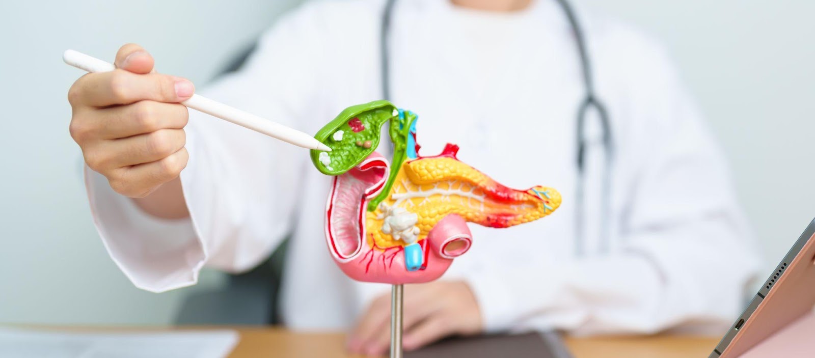
Pigment stones only account for a very small percentage of gallstones. These stones are formed as a result of high bilirubin levels in the bile, leading to crystallisation and formation of pigment stones. The high bilirubin levels are due to the breakdown of red blood cells and hence occurring in individuals with red blood cell disorders like thalassemia, sickle cell anaemia and hereditary spherocytosis. Individuals with pigment stones tend to be much younger in their 20s to early 30s.
Nestled under the right side of the liver, the gallbladder sits in a shallow depression in the liver called the gallbladder fossa. This area lies in the upper right abdomen, protected by the lower rib cage. Its position explains many symptoms experienced during gallbladder disease.
Because the gallbladder shares nerve pathways with the diaphragm, shoulder and back, the pain often spreads beyond the abdomen. Many people describe discomfort as:
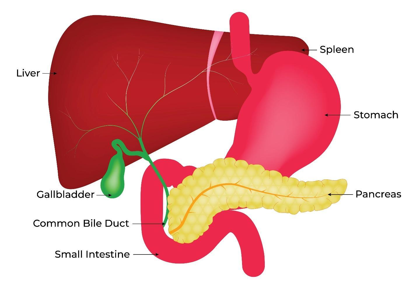
Although small, the gallbladder is divided into 3 parts, each with a specific function in storing and releasing bile. The structure includes:
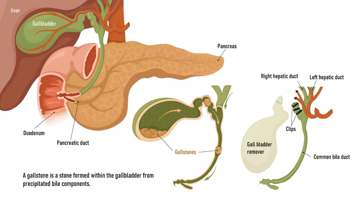
The gallbladder’s blood vessels and nerve supply are essential not only for its function but also for safe surgical removal. This consists of:
The gallbladder does not work alone. It is part of a connected system of ducts known as the biliary tree, which carries bile from the liver to the small intestine, which includes:
A clear understanding of the gallbladder anatomy helps explain many common issues, including:
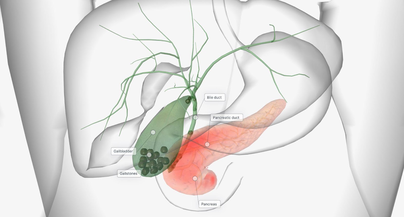
The anatomical connections between the liver, pancreas and intestinal tract mean that even a single stone in the wrong place can disrupt multiple digestive processes.
Like other organs, the gallbladder is susceptible to conditions that develop over time and may disrupt normal bile storage and release.
Condition | What It Means | Typical Symptoms |
Gallstones (Cholelithiasis) | Hardened deposits in the gallbladder | Pain, nausea, bloating, discomfort after meals |
Gallbladder Inflammation (Cholecystitis) | Irritation or infection of the gallbladder | Persistent pain, fever, vomiting |
Biliary Colic | Intermittent pain from gallstone blockage | Sudden pain after meals, nausea |
Bile Duct Stones (Choledocholithiasis) | Gallstones lodged in the bile duct | Jaundice, dark urine, abdominal pain |
Gallbladder Polyps | Tissue growths on the gallbladder wall | Usually no symptoms, usually incidentally found on imaging. Occasionally can have symptoms similar to gallstones. Large polyps >1cm in size should be removed due to risk of malignant change |
When the gallbladder becomes a source of repeated pain, infection or complications, surgical removal is often recommended. Most procedures are performed using minimally invasive techniques, such as laparoscopic or single-incision surgery, allowing faster recovery and less postoperative discomfort.
During surgery, the surgeon identifies the cystic duct and cystic artery using a safety technique known as the Critical View of Safety. This method ensures the correct structures are separated before removal, significantly reducing the risk of injury to the bile ducts.
Life without a gallbladder is entirely normal for most people. Bile continues to flow from the liver directly into the intestine and digestion typically adjusts over time. Some individuals may experience temporary changes in bowel habits, but these usually improve within weeks.
Although small, the gallbladder plays an important role in storing and delivering bile for digestion. Its anatomy, from its position under the liver to its connection with the bile ducts, explains many symptoms seen in gallstone disease. Recognising how this organ functions can help you understand your condition better, interpret scan results more confidently and make informed treatment decisions.
If you experience persistent upper abdominal pain, nausea after meals or jaundice, we encourage you to book a consultation with Dr Aaron Poh. Medical evaluation is recommended to identify the cause early, prevent complications and receive timely intervention.

Dr Aaron Poh obtained his M.B.B.S (Bachelor of Medicine & Bachelor of Surgery) from the National University of Singapore in 2004. He then entered the MOH General Surgical training programme in 2006 and obtained his Fellowship from the Royal College of Surgeons of Edinburgh (F.R.C.S Edinburgh) in 2015. He was also accepted into the Colorectal Fellowship programme in the Queen Elizabeth Hospital in Birmingham, UK in 2014. His special interest is in minimally invasive colorectal cancer surgery and exenterative surgery for advanced cancers.


Patients are advised to have a consultation with our Surgical Specialist, Dr Aaron Poh to first understand your condition and concerns. After referring you to the appropriate screening tests, he will advise you of your diagnosis and treatment options after the tests and scopes


Copyright © Gallstones Doctor Singapore by Alpine Surgical Practice | Terms & Conditions

Copyright © Gallstones Doctor Singapore by Alpine Surgical Practice | Terms & Conditions